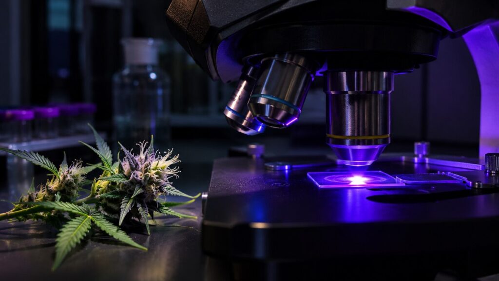
There is a landscape on the surface of a cannabis flower that almost disappears in a photograph of the whole plant. Magnify a small patch and it fills with translucent heads, some perched on stalks, others close to the surface. Under carefully chosen illumination, the heads can give off light of their own. The startling colors in the resulting image tell a story about plant development, provided you know what the microscope actually measured.
Where the light comes from
A specimen examined by ordinary reflected light sends some of the illumination back toward the observer. In fluorescence microscopy a substance absorbs light and emits light at another wavelength. Filters help separate that emitted light from the illumination. Sometimes a scientist adds a fluorescent stain so that a particular structure stands out. Plant tissue can also contain substances that fluoresce without an added label. That property is called autofluorescence.
In a microscopy study of cannabis glandular trichomes, investigators made use of this natural signal. Glandular trichomes are specialized structures where the plant produces and accumulates compounds including cannabinoids and terpenes. The scientists combined images with information about the cells in the glands and the substances the glands contained. A color difference became interesting because there were independent observations against which to check it.
A blue head is an observation
Stalked glandular heads in the study showed blue autofluorescence associated with high cannabinoid levels. That finding has a narrower meaning than a photograph sometimes suggests. A patch of blue in a published micrograph belongs to a particular preparation, illumination and detection method. A camera looking at a flower on a windowsill is collecting a different signal. Even on the same microscope, an exposure setting can make a dim object look bright. A guide to quantitative fluorescence microscopy explains why those settings matter when the goal is to measure intensity.
Imagine photographing two identical stars, once with a short exposure and once with a long one. Their brightness in the resulting pictures would differ without either star having changed. Plant microscopists face a similar problem when comparing tissues. Color channels, exposure and the ways researchers display an image are part of the evidence. The accompanying caption can be as important as the picture.
Is a short gland a young tall gland?
A collection of photographs can resemble a family album in which nobody wrote the dates. A short structure and a tall one might be different types, or one might develop into the other. The researchers examined features beyond height, including the number of secretory cells and changes seen as flowers matured. Several observations together supported a developmental relationship between stalked glands and precursors that looked superficially like structures without stalks.
The distinction matters when counting. Treat every short gland as a single category and you may combine structures with different histories. A later study using image analysis to quantify cannabis trichomes approached the practical problem of measuring many glands. Whatever method is used, the definition of a gland type has to survive a change of angle, focus or stage of growth.
What remains outside the picture
The glands help explain why scientists interested in smell examine the flower surface so closely. But the move from an image to a scent requires further evidence. Chemical analysis of volatile sulfur compounds addresses molecules that may have a large sensory effect even in tiny quantities. A luminous head in a micrograph cannot tell the viewer what a person standing beside the plant would smell.
Look again at the apparent forest of stalks. Height, color, cell number and chemical composition are four descriptions of the same piece of living tissue. They can agree in informative ways, as they did in the microscopy study. The point of measuring all four is that no one of them can simply stand in for the others.